Feline Stomatitis:
One of the most painful and frustrating conditions cats can develop is feline stomatitis. The cause is unknown, but appears to be a severe reaction to plaque and the tooth structure itself, or the basement membrane of the periodontal tissues. Clinical signs include severe chronic gingivitis, with or without the following: faucitis, pharyngitis, or palatitis (inflammation of the throat or roof of the mouth). Other signs include excessive salivation, reluctance to eat, bleeding gum tissue, extreme oral sensitivity, and weight loss or anorexia. Diagnosis is based on clinical signs and biopsy of the affected gingival tissue. The histopathology usually finds abundant lymphocytes, plasmacytes and occasional neutrophils. Although many organisms have been cultured or found in affected cat’s mouths, none has been proven to be the cause. As such, oral culture and sensitivity or viral isolation has been of little benefit.
Case management of a cat with stomatitis should include CBC, full chemistry profile, urinalysis, T4 and free T4, FeLV and FIV testing, and gingival biopsy. Cleaning the teeth, homecare/brushing, oral antibiotics and corticosteroids are helpful initially, but their effectiveness for treatment usually wanes within 3-6 months. The only treatment thus far shown to have long-term results without the need further medication is either caudal or full mouth dental extraction. In the only study to report long term results of caudal or full mouth extractions, 60% had significant improvement, another 20% had some improvement, and a final 20% had little to no clinical improvement in the gingivitis, but we have seen that most seem to be more comfortable. Dental radiographs are essential when performing these extractions to ensure the entire root of every extracted tooth is removed. Post-operatively, antibiotics are given for 14-28 days. Pain management is paramount in these patients and is accomplished with pre-anesthetic opioid administration, intraoperative local anesthetics (Bupivacaine), and postoperative NSAID and opioid given orally for at least 5-7 days. For those patients with anorexia prior to presentation, nutritional support via esophagostomy or gastrostomy tube may be warranted either pre or post-operatively until eating well again. Re-evaluation at one month should show some improvement, and further follow-up at three months should be indicative of success of treatment. If there is little to no response, the remainder of the teeth may need to be extracted or dental radiographs taken to ensure all tooth/root remnants have been removed and that there are no areas of reactive alveolar bone borders.
The question that consistently arises is “How can these patients eat without their teeth?” The fact of the matter is that the one’s who respond favorably do tremendously better without the oral pain and chronic infection eating soft food than they ever did before, and those who still have inflammation seem to be more comfortable. Many continue to eat dry kibble even without teeth!
'The Pet Dentist' - Location, Map, Directions. Offices serving greater Tampa Bay, Clearwater, St Petersburg, Brandon & Bradenton :


Canine Stomatitis: (Chronic Ulcerative Paradental Stomatitis)
Similar to cats, dogs too can have stomatitis. In dogs, the underlying etiology seems to be related to a severe reaction to plaque on the teeth surfaces. The hallmark clinical sign in these cases is “kissing” ulcers of the cheek and lip mucosa over the teeth covered with a soft creamy plaque. There is usually severe halitosis, ulcerations on the lateral margins of the tongue, and extreme sensitivity within the oral cavity. Some dogs will stop eating, but most continue reluctantly. Histopathology (biopsy) of the oral ulcerations usually results in chronic active inflammation with mucosal ulceration.
Early in the course of disease, cleaning the teeth can be beneficial if the owners can brush the teeth. This is usually difficult because the pets are sensitive around their mouths and as plaque accumulates, the ulcers tend to return within 4-6 weeks. Adjunctive modalities include adding antiseptics to the drinking water, pulse antibiotic therapy, oral chlorhexidine rinses, or barrier sealants to help slow/reduce plaque accumulation. Over time, the effectiveness of these efforts tend to diminish and more aggressive therapy is needed. In advancing cases, removal of the source of plaque accumulation, i.e. the teeth, usually results in resolution of the oral ulcerations. Although there are no studies to show response to extraction therapy, my experience has been that these are more responsive than cats and significant relief can be obtained from partial or full mouth dental extractions.
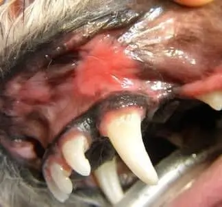
“Kissing ulcers” where the oral mucosa touches a plaque laden tooth is typically of a case of canine ulcerative stomatitis.